Pinkeye
Pinkeye (Infectious Bovine Keratoconjunctivitis)
Image courtesy of Beef Magazine
The term “pinkeye” is often used to describe a variety of eye diseases affecting cattle, sheep, and goats. However, in cattle, the term most commonly refers to infectious bovine keratoconjunctivitis (IBK), a contagious eye disease characterized by central corneal ulceration and the development of a cloudy or bluish appearance in the eye.
CAUSE
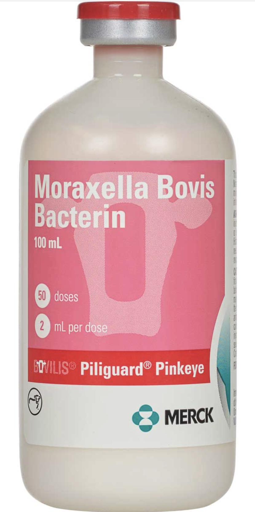
Pinkeye in cattle has traditionally been associated with the bacterium Moraxella bovis. However, more recent research has shown that other organisms, including Moraxella ovis and Mycoplasma species, can also contribute to the disease. This is an important consideration when developing prevention strategies and selecting vaccines.
Moraxella bovis is primarily spread by face flies. The bacterium attaches to the surface of the eye using hair-like structures called pili. Once attached, it produces toxins that damage and destroy cells on the surface of the cornea. This damage leads to the formation of the characteristic central corneal ulcer, which is the hallmark lesion of infectious bovine keratoconjunctivitis (IBK), or "true" pinkeye in cattle.
CLINICAL SIGNS
Pinkeye cases typically begin appearing in late spring and early summer as fly populations increase, with cases often continuing through late summer and into the fall. The severity of pinkeye can vary considerably, ranging from mild irritation that resolves on its own to severe infections that can permanently damage or even rupture the eye.
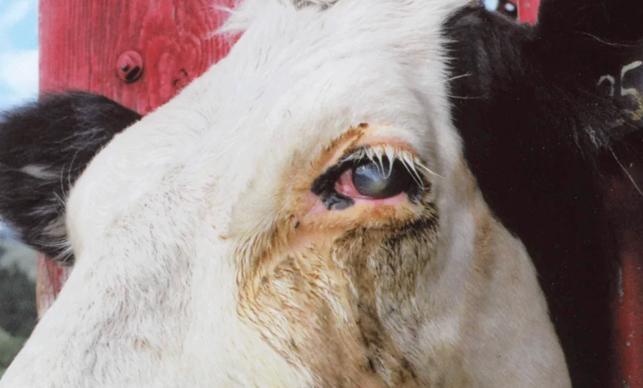
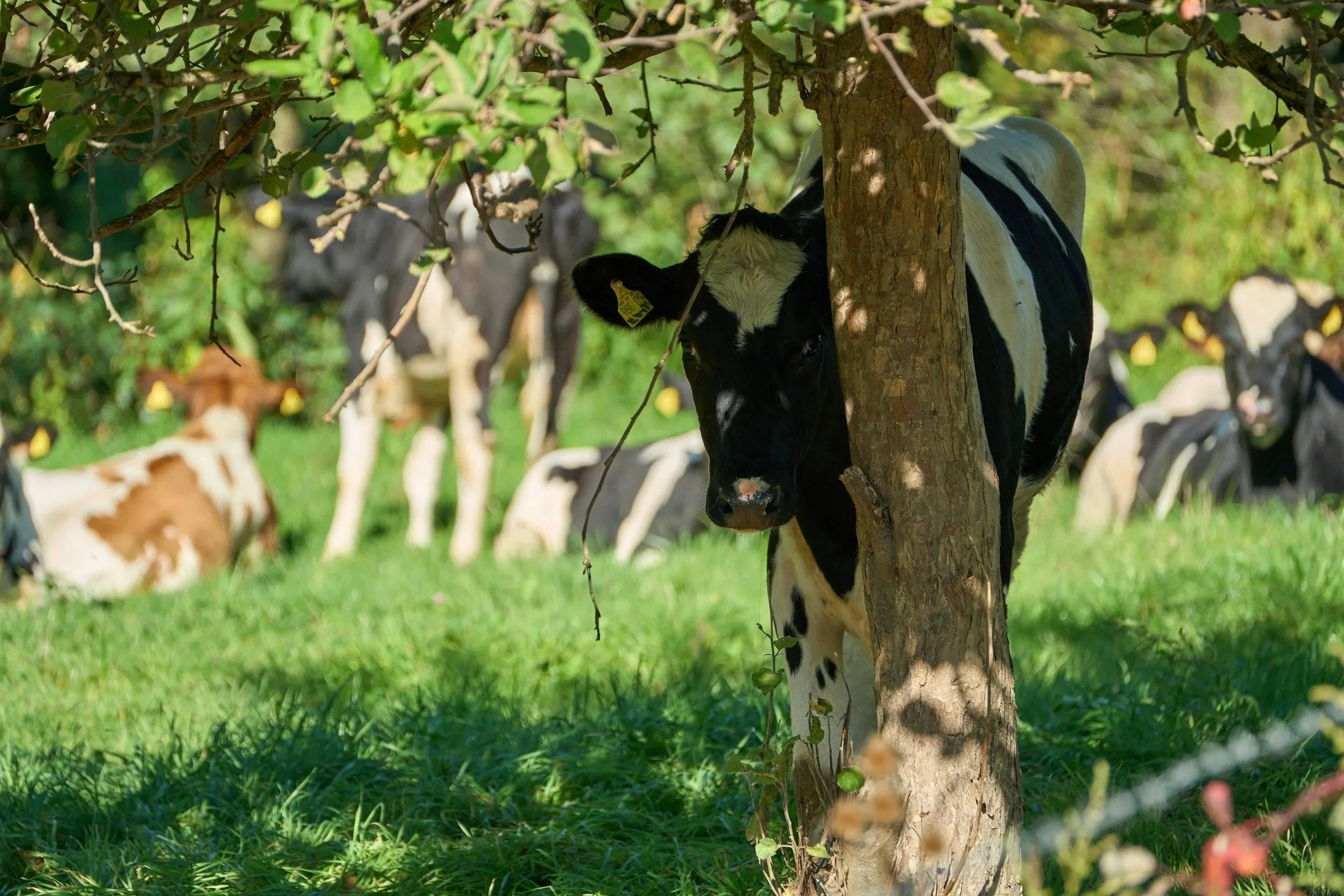
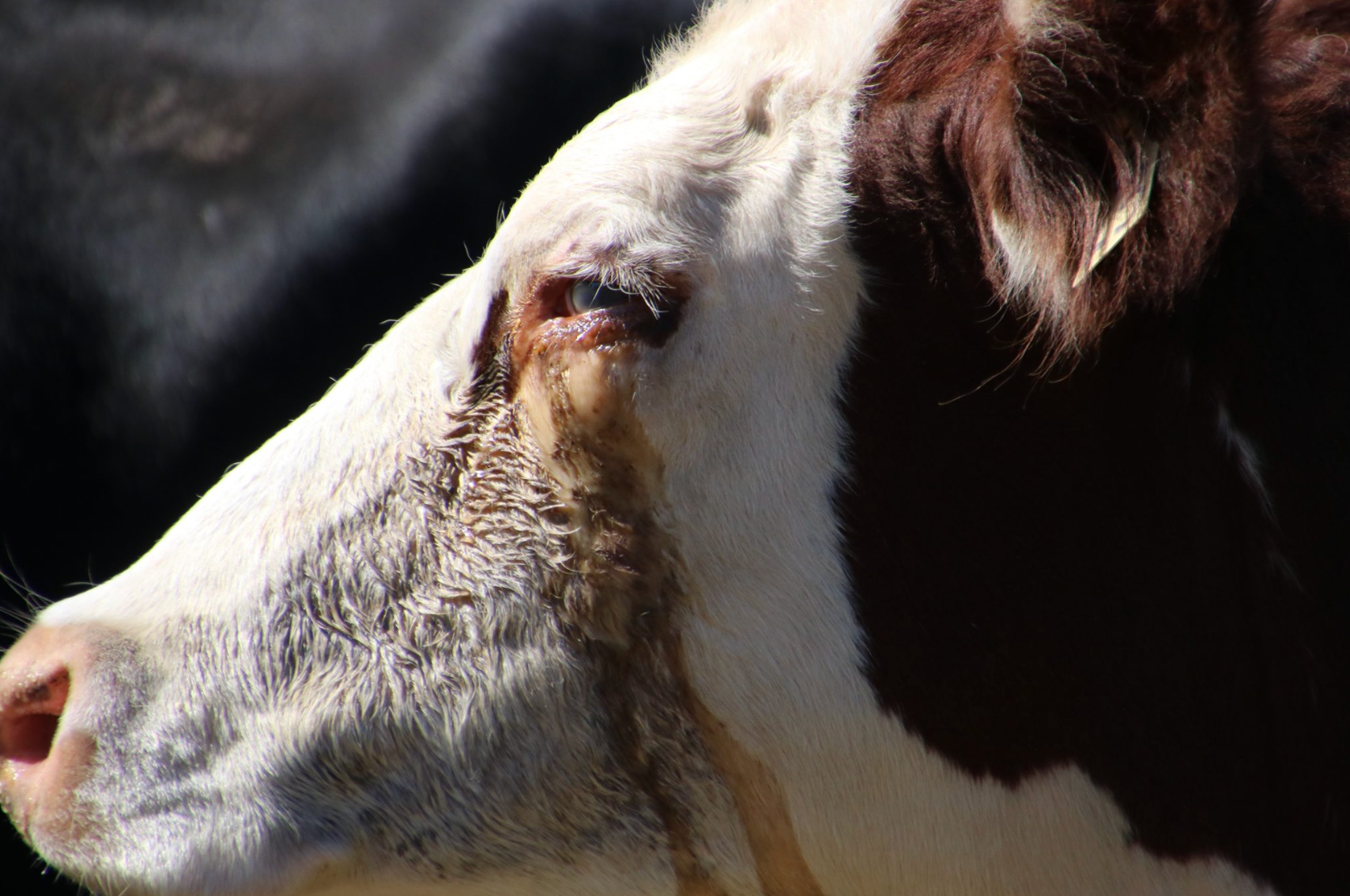
The earliest signs of pinkeye are often subtle and can be easily overlooked. In most cases, only one eye is affected initially, although both eyes may become involved. Early symptoms include excessive tearing, mild squinting, and redness around the eyelids. Because corneal ulcers are painful and make the eye sensitive to light, affected cattle may seek shade, separate themselves from the herd, or spend less time grazing during bright, sunny conditions.
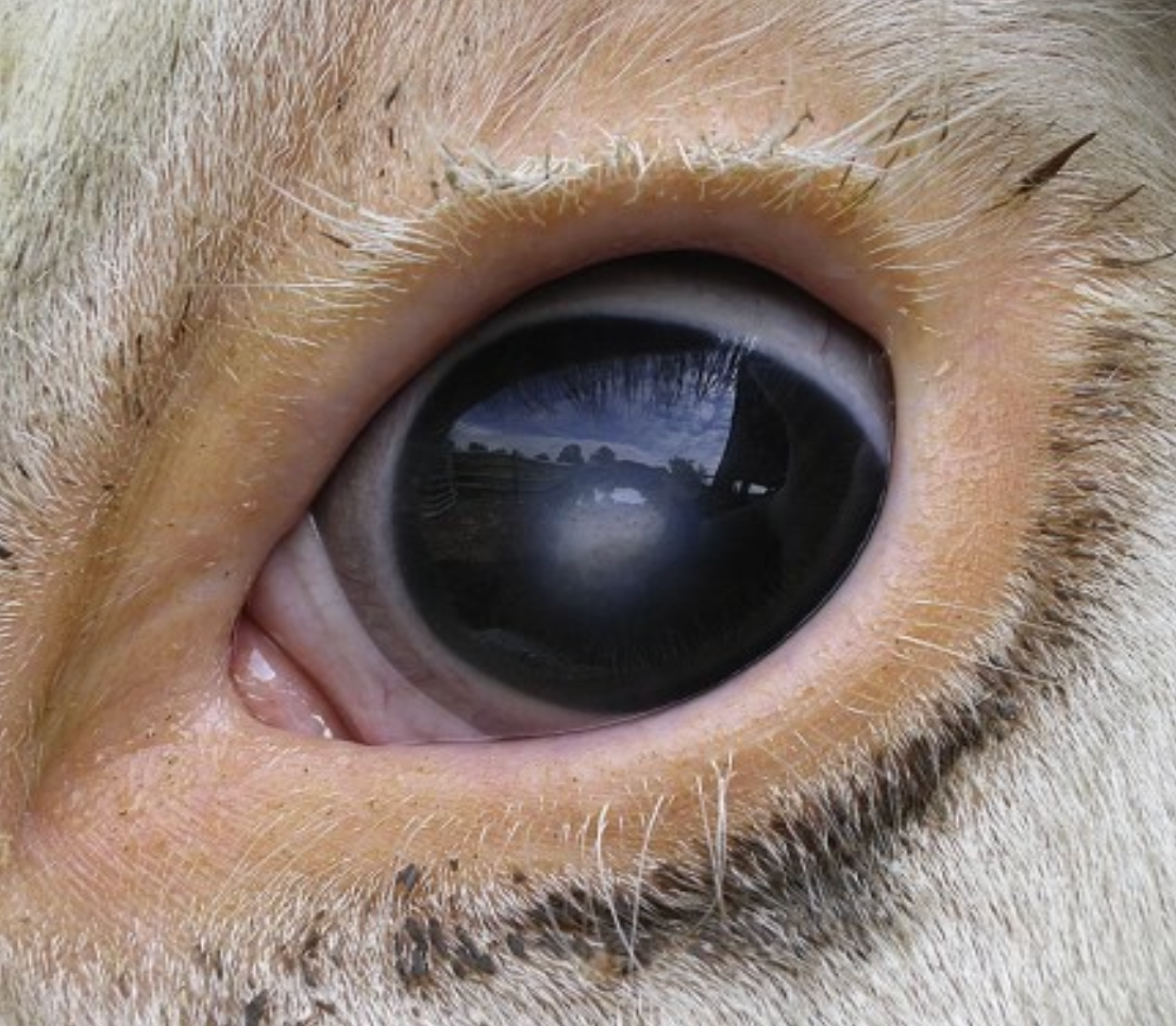
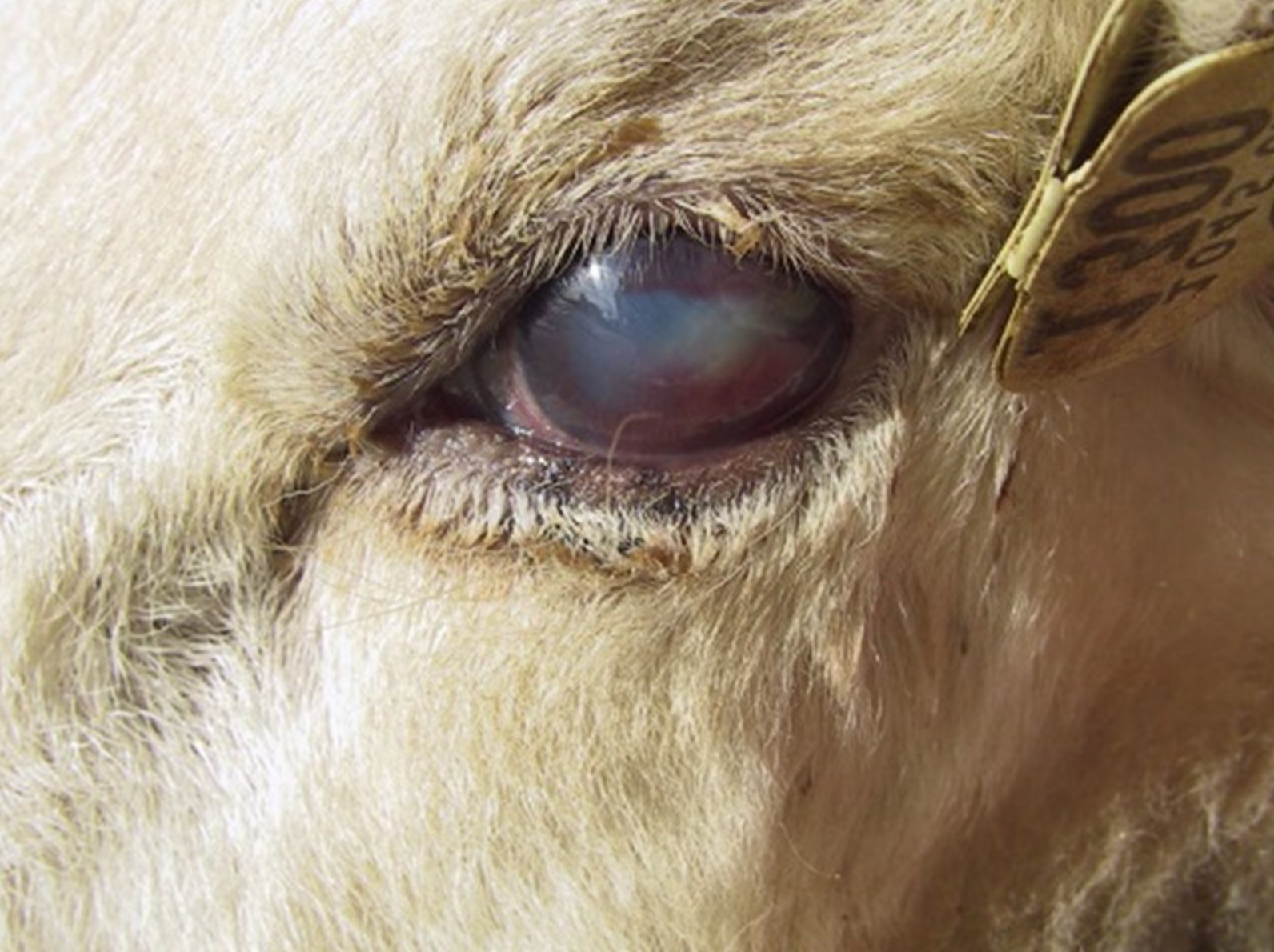
Mild Pinkeye - Excessive Tearing and Blue Eye
Image courtesy of Auburn Extension
Mild cases may resolve without treatment, but some progress to a more moderate stage of disease. As the infection advances, a small ulcer develops in the center of the cornea, appearing as a distinct white spot. The cornea may also become cloudy or develop a bluish appearance due to swelling (corneal edema). These moderate cases are often the stage at which producers first notice a problem and seek veterinary assistance. Although some moderate cases may eventually heal on their own, veterinary evaluation is generally recommended because corneal ulcers are painful and can worsen if left untreated.
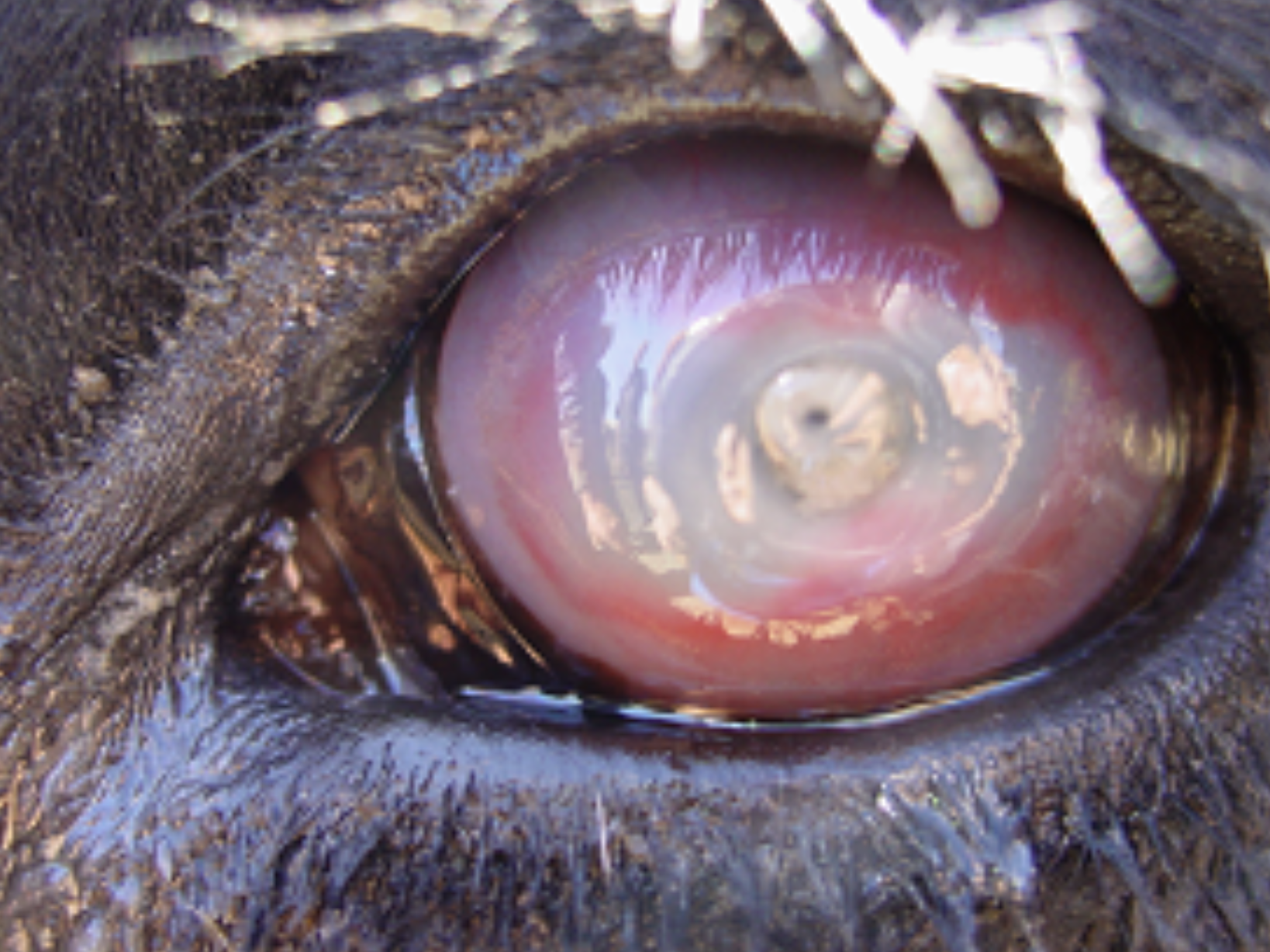
Severe Pinkeye - Melting Corneal Ulcer
Image courtesy of NADIS Animal Health
Severe cases are less common but can have serious consequences, including permanent vision loss. In these animals, the corneal ulcer deepens and the cornea may take on a soft, gelatinous appearance, a condition known as a “melting ulcer.” If the ulcer continues to progress, it can penetrate the full thickness of the cornea and cause the eye to rupture. Because these cases represent a medical emergency, affected cattle should be examined by a veterinarian as soon as possible.
OUTCOME
Corneal Scar
Image courtesy of University of Wisconsin-Madison Extension
It is important to recognize that pinkeye can vary greatly in both severity and outcome. Not every animal exposed to the bacteria will develop clinical signs, and not every case will progress to corneal scarring or loss of vision. In fact, most cases of pinkeye resolve without treatment. Studies have reported that approximately 98% of affected cattle recover spontaneously, and permanent blindness occurs in only about 1 out of every 50 cases.
Even when a corneal scar develops, the long-term impact on vision is often limited. These scars typically appear as a white spot in the center of the eye after the infection has resolved and are not usually associated with ongoing tearing or squinting. Although the scar may partially obstruct vision, most cattle are able to see around it and function normally with few noticeable problems.
However, vision loss is not the only consequence of pinkeye. The disease is painful, particularly when a corneal ulcer is present. Pain and light sensitivity can cause affected cattle to spend less time grazing, which may reduce weight gain in growing animals and decrease milk production in lactating cattle. In addition, visible eye damage, including corneal scars and permanently shrunken or scarred eyes, can reduce an animal’s market value and negatively affect overall herd productivity.
TREATMENT
Any animal suspected of having pinkeye should be evaluated by a veterinarian. Cases identified early in the disease process generally respond best to treatment, while moderate to severe cases often require more intensive care to prevent permanent damage to the eye. In cattle, two injectable antibiotics are specifically labeled for the treatment of pinkeye: oxytetracycline (LA-200/300®) and tulathromycin (Draxxin®). However, by the time many cases are recognized, a corneal ulcer has already formed and significant tissue damage may have occurred. In these situations, antibiotic treatment may have limited ability to reverse the existing damage, although it can still help control the infection. Pain-relieving medications, such as meloxicam or flunixin meglumine (Banamine®), are often used to improve comfort and reduce inflammation.
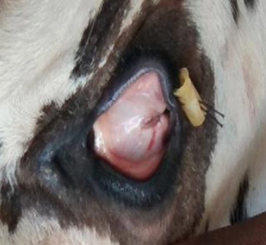
Third Eyelid Flap
Image courtsey of The Pharma Journal
Topical antibiotic ointment formulations, such as Oxytetracycline Hydrochloride and Polymyxin B Sulfate Ophthalmic Ointment (Terramycin®), are also frequently used. While they can be effective, they generally require application at least twice daily for optimal results, which is often impractical in many cattle operations. Over-the-counter products containing hypochlorous acid, such as Vetricyn, are marketed for pinkeye treatment and work by reducing bacterial contamination on the surface of the eye. However, these products also require frequent application and do not address the significant pain and inflammation associated with corneal ulcers.
Protecting the affected eye is another important component of treatment. Ulcerated eyes are extremely sensitive to light, so covering the eye can provide significant pain relief. Eye coverings may also help reduce bacterial transmission, although pinkeye is highly contagious and herd exposure has often already occurred by the time moderate or severe cases are detected. Commercial eye patches are commonly used but can be difficult to keep in place. An alternative approach is a third-eyelid flap, a procedure performed by your veterinarian in which the third eyelid is temporarily sutured over the cornea to protect it during healing. Because absorbable suture material is typically used, the flap does not require removal and often remains in place longer than a conventional eye patch.
It’s important to consider drug withdrawal periods when treating cattle for pinkeye. These are listed on the product label and indicate how long it takes for drug residues to drop to safe, legal levels before meat or milk from treated animals can enter the food supply. Both injectable and topical antibiotics require a prescription, so you should consult your veterinarian for appropriate treatment options and herd management guidance.
PREVENTION
Effective pinkeye prevention focuses on two main goals: reducing cattle exposure to the bacteria that cause the disease and minimizing factors that allow those bacteria to infect the eye.
Pinkeye is a highly contagious disease, but exposure alone does not guarantee that an animal will develop clinical signs. The risk and severity of infection are influenced by several factors, including the animal's overall health, immune function, genetics, and environmental conditions. Understanding and managing these risk factors is the foundation of an effective pinkeye prevention program.
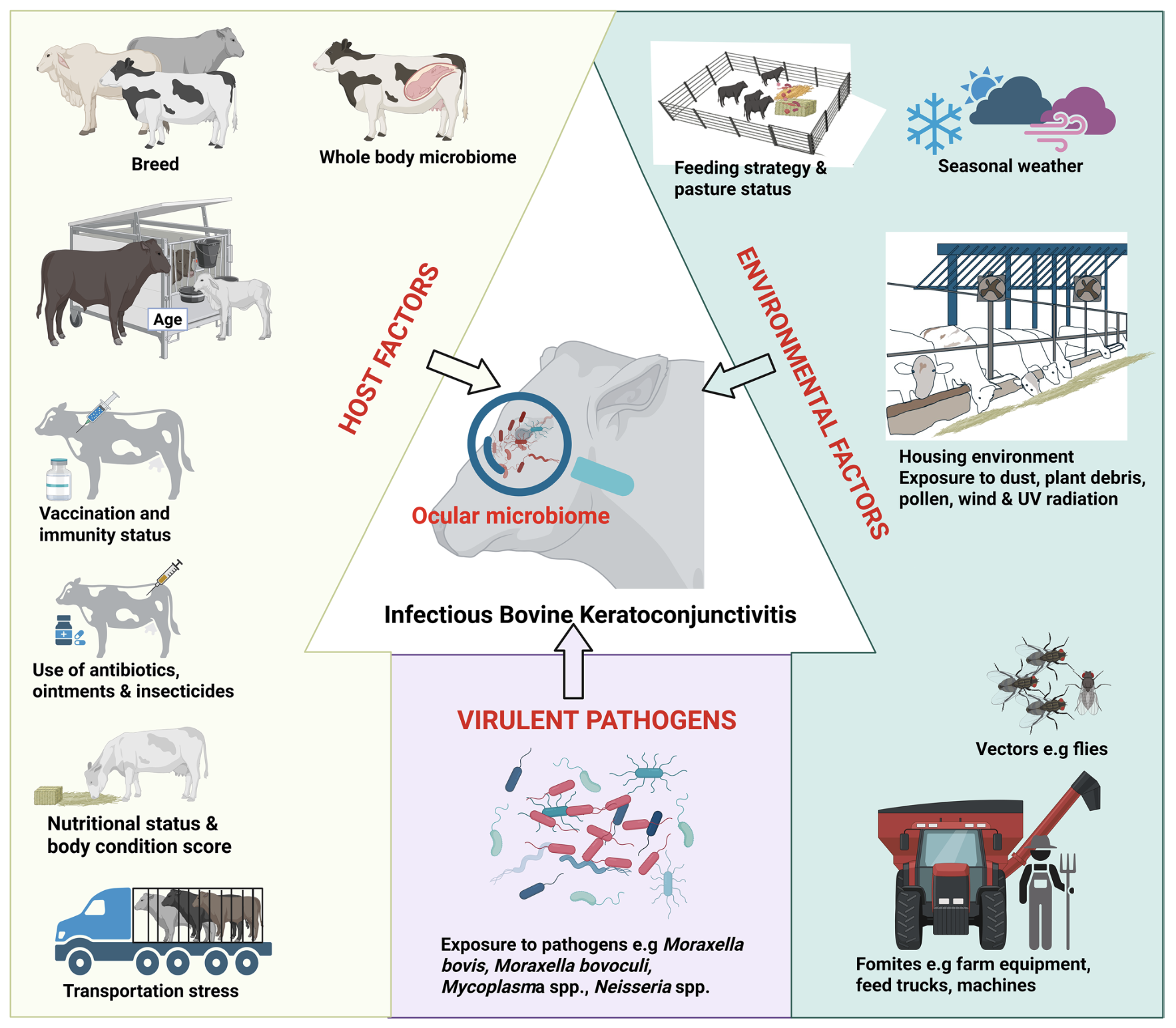
Factors Contributing to Development of Pinkeye
Diagram courtesy of Kilama, et al.
Isolating Cases
Early detection and isolation of affected animals are important for reducing the spread of pinkeye within the herd. Take the opportunity to closely examine your cattle’s eyes each time they are handled or processed through the chute. Animals treated in the early stages of infection typically respond better to treatment and are less likely to develop permanent corneal scarring. Promptly isolating infected animals and protecting the affected eye with an eye patch or third eyelid flap can also help reduce transmission to other cattle.
Preventing Cornea Damage

Anatom of the Eye
Diagram courtesy of Kilama, et al.
For pinkeye to develop, the bacteria must penetrate the cornea. As a result, anything that irritates or damages the eye can increase an animal’s risk of infection. Healthy eyes have natural defense mechanisms that help prevent disease, but scratches or irritation to the cornea can compromise these defenses, allowing bacteria to enter and cause infection.
Several environmental factors can contribute to corneal damage. Exposure to ultraviolet (UV) light can injure the cornea, so providing adequate shade for cattle can help reduce the risk of pinkeye. Dust and debris can also irritate or scratch the eye, making windbreaks a useful management tool. Additionally, tall grasses and weeds can cause eye trauma, so regular mowing of pastures may help prevent injuries.
Overall, management practices that minimize eye irritation and trauma can play an important role in reducing the incidence of pinkeye in cattle.
Fly Control
Pinkeye causing bacteria is spread through flies so effective fly control is another corner stone of pinkeye prevention. Pinkeye is spread by the face fly (Muscaautumnalis), which is attracted to the normal eye and nasal secretions cattle produce.
Insecticide ear tags can help control face flies because they provide protection close to the cattle’s head and eyes. However, fly populations can develop resistance to insecticides overtime, so rotating the active ingredient used in ear tags is recommended. You also want to chose a product that is labeled specifically for face fly control. Most tags are labeled for horn flies which are usually not responsible for the spread of pinkeye.
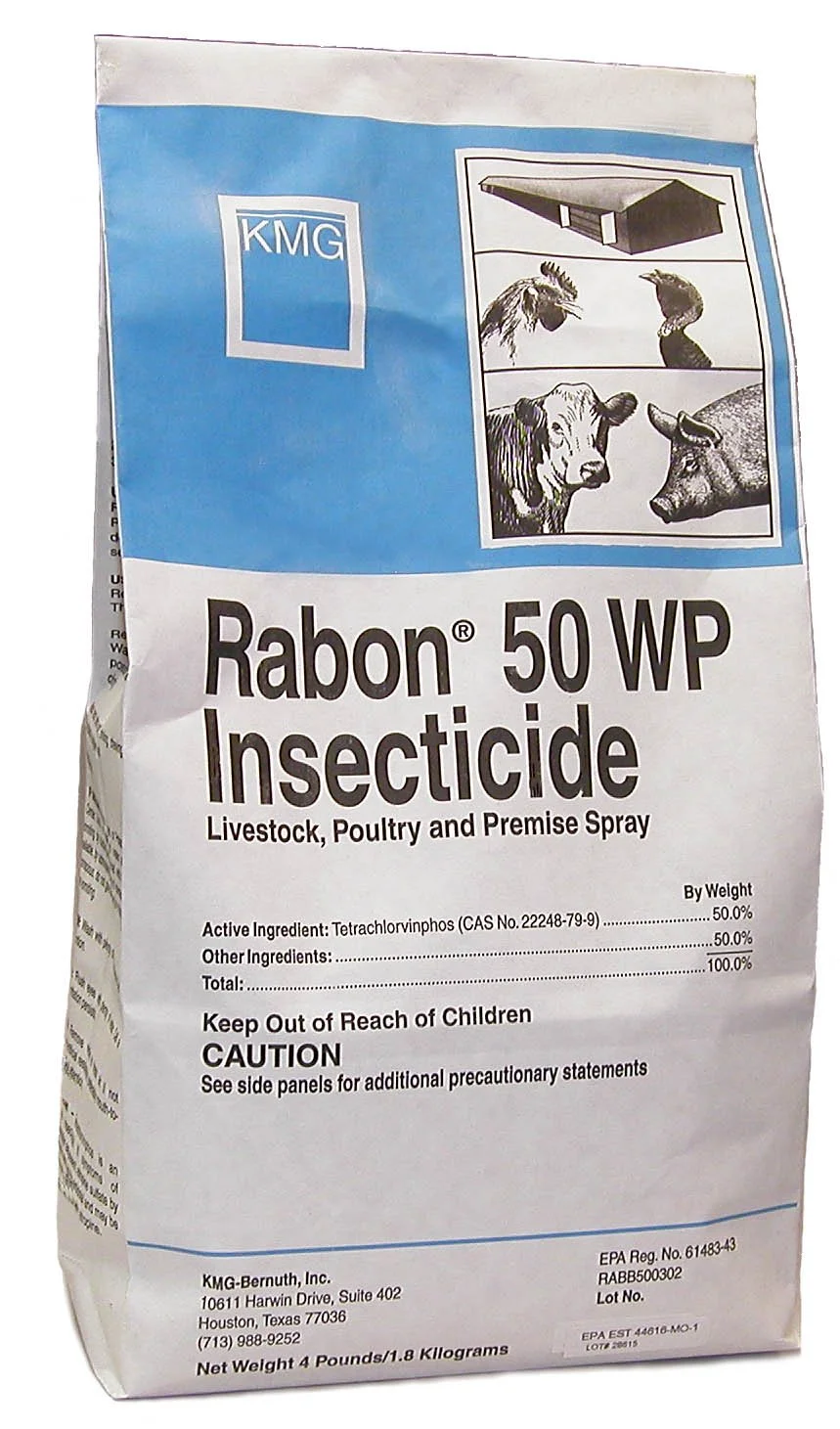
Feed through fly larvicide can also be useful since face flies only lay eggs in freshly deposited manure. Tetrachlorvinphos (Rabon®) will kill the larvae deposited in the fresh manure, but it is most effective when it is started early before fly season, the animal receives the required dose everyday, and it is fed through the entire fly season. Fly sprays containing organophosphates or permethrin may provide additional control, but their effectiveness is typically short-lived and requires frequent reapplication.
Since face flies are most active during daylight hours, grazing cattle at night may help reduce fly exposure. It is also important to understand that face flies lay their eggs in freshly deposited manure, typically within minutes after it is dropped. As a result, removing old manure from pastures will not significantly reduce face fly populations, although it may help control other fly species.
Nutrition

An animal’s overall health and immune function also influence its susceptibility to pinkeye and the severity of infection. Stress, poor nutrition, and other factors that compromise the immune system can increase the risk of disease. Adequate trace mineral supplementation (especially copper, zinc, iodine, and selenium) is essential for maintaining a healthy immune response. More information on trace mineral supplementation can be found here – Guide to Trace Minerals.
Vitamin A is especially important for eye health because it helps maintain the integrity of the corneal and conjunctival surfaces, which serve as the eye’s first line of defense against bacteria and viruses. Adequate vitamin A levels can help reduce the risk of eye infections, respiratory disease, and other infectious conditions. Herds experiencing frequent or severe cases of pinkeye should have their nutrition program evaluated, with particular attention to trace mineral and vitamin supplementation.
Genetics
Genetics can also influence an animal’s risk of developing pinkeye. In general, Bos taurus breeds, such as Herefords and Angus, tend to be more susceptible than Bos indicus breeds, such as Brahmans and Zebus. Cattle with white pigmentation around the eyes are also at greater risk than cattle with darker facial pigmentation, likely because increased tearing attracts more face flies. Age is another important factor, with most cases occurring in younger cattle. Animals under two years of age are generally more susceptible than mature cattle.
Vaccination
Two types of vaccines are available for pinkeye prevention: commercial vaccines and autogenous vaccines. Commercial vaccines are mass-produced products, while autogenous vaccines are custom-made using bacterial strains isolated from affected animals within a specific herd.
Most commercial pinkeye vaccines are designed to target Moraxella bovis, the bacterium traditionally associated with infectious bovine keratoconjunctivitis (IBK). However, research has shown that pinkeye can be caused by multiple bacterial species, including Moraxella ovis and Mycoplasma species. In addition, considerable variation exists among M. bovis strains themselves. Because of this diversity, the effectiveness of commercial vaccines can be inconsistent. Clinical studies have generally demonstrated limited or highly variable protection from commercially available pinkeye vaccines.
Autogenous vaccines are developed from bacterial isolates collected directly from affected animals in a herd and are intended to provide more targeted protection. While this approach is theoretically appealing, research has also shown mixed results, with vaccine effectiveness varying widely between herds and outbreaks. One challenge is that the bacterial strains responsible for pinkeye can change from year to year, meaning an autogenous vaccine developed for a herd one season may not provide adequate protection in subsequent years. Recent reviews of the scientific literature, including those published in Veterinary Clinics of North America Veterinary Clinics: Food Animal Practice, have found that both commercial and autogenous vaccines generally provide poor protection against pinkeye.
Timing is also an important consideration. Following vaccination, the immune system requires several weeks to develop a protective response. To maximize effectiveness, pinkeye vaccines should be administered approximately 3 to 5 weeks before the start of fly season. In Northern California, this typically means vaccinating cattle in March before face fly populations begin to increase.
For herds experiencing frequent pinkeye outbreaks or cases that respond poorly to treatment, diagnostic testing may be beneficial. Your veterinarian can collect samples for culture and other laboratory testing to identify the specific organisms involved. This information can help guide treatment decisions and determine whether an autogenous vaccine or other herd-specific prevention strategies may be warranted.
WHAT IS NOT “PINKEYE”
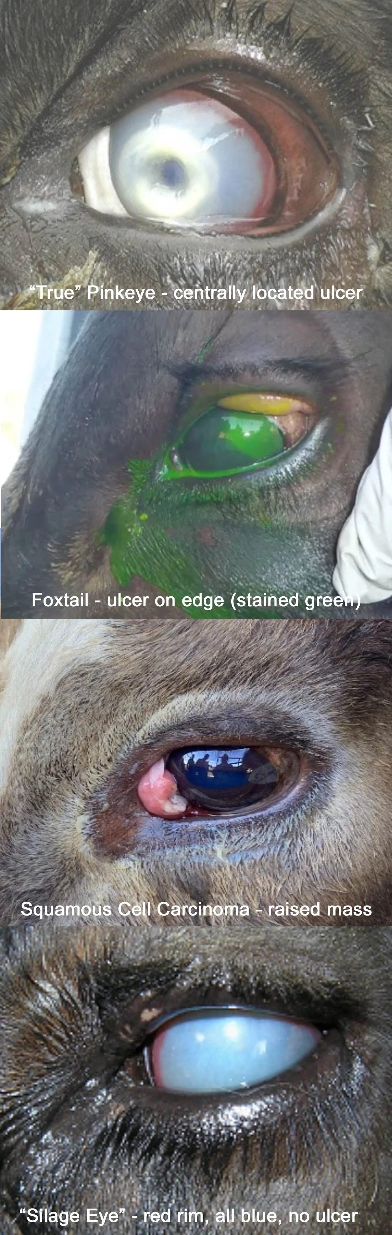
One of the most common reasons pinkeye treatments fail is that the condition being treated is not actually pinkeye. Accurately distinguishing pinkeye from other eye diseases is critical, as it can significantly affect whether an animal retains normal vision.
Foreign Body (Grass, Foxtail, Debris)
A key feature of pinkeye (infectious bovine keratoconjunctivitis) is a centrally located corneal ulcer. If the ulcer is located on the edge of the cornea instead of the center, this more commonly indicates a foreign body such as grass awns or foxtails.
Another clue is that foreign body injuries are often isolated cases affecting a single animal, whereas pinkeye is highly contagious and typically results in multiple affected animals within a herd. However, it is important to note that any corneal scratch or injury can predispose the eye to secondary pinkeye infection because the damaged cornea allows bacteria to enter. For this reason, animals with removed foreign material should still be monitored closely until the eye has fully healed.
Squamous Cell Carcinoma (“Cancer Eye”)
Pinkeye does not cause masses or tumor-like growths. If a raised, proliferative lesion is present on the eye—especially in white-faced cattle—this is more consistent with squamous cell carcinoma (“cancer eye”). These cases should be evaluated by a veterinarian for diagnosis and treatment options.
Listeria monocytogenes (“Silage Eye” or “Winter Pinkeye”)
Listeria monocytogenes is a bacterium commonly found in soil and spoiled or moldy feed, and cases are often associated with silage feeding. It typically causes inflammation involving the entire eye and is more commonly seen during winter months. This seasonal pattern, along with the diffuse nature of the inflammation, helps distinguish it from pinkeye, which is most common in summer and characterized by a central corneal ulcer.
Viral Diseases
Several viral infections can also cause eye lesions in cattle. Infectious bovine rhinotracheitis (IBR), a herpesvirus, typically causes redness and inflammation at the junction between the cornea and sclera (the clear and white parts of the eye). Other viral diseases, including malignant catarrhal fever (MCF) and bovine viral diarrhea virus (BVDV), can also occasionally lead to eye involvement.
In general, viral eye disease tends to cause more diffuse irritation rather than a well-defined central corneal ulcer. The presence of a central ulcer is therefore much more suggestive of true pinkeye.
Consult Your Veterinarian
Because the eye has a limited number of ways it can respond to injury or disease, different conditions can look very similar in the early stages. A veterinary examination is often necessary to make an accurate diagnosis and ensure appropriate treatment.
PINKEYE IN GOATS AND SHEEP
“Pinkeye” in a sheep
Image courtesy of NADIS Animal Health
Goats and sheep can develop pinkeye, but it differs from the form seen in cattle. In small ruminants, the disease is caused by organisms such as Mycoplasma, Chlamydia, and Moraxella ovis. It typically causes inflammation of the entire eye, often affects both eyes, and does not usually produce a central corneal ulcer.
Because it is highly contagious, multiple animals are usually affected. If only one goat or sheep shows signs, the eye should be closely examined for a foreign body such as grass or a foxtail. If pinkeye is suspected in a flock or herd, consult your veterinarian for diagnosis and treatment recommendations.
KEY TAKEAWAYS
Pinkeye is the most common and economically important eye disease in cattle. It mainly affects animal well-being and productivity, leading to losses from reduced weight gain, lower milk production, and treatment costs.
Even though it spreads easily, most cattle that get pinkeye only have mild signs that go away on their own and don’t result in vision loss.
Catching cases early and accurately is important for getting the best outcome for the animal and helping prevent it from spreading to others.
REFERENCES
Jones, M. Course 42: Pinkeye in Cattle. Large Animal Continuing Education.
Kilama, J., Islam, Md. S., & Amat, S. (2025). Bovine ocular microbiome: The Next Frontier in managing pinkeye in cattle. Animal Microbiome, 7(1). https://doi.org/10.1186/s42523-025-00425-9
O’Connor, A. Infectious Bovine Keratoconjunctivitis: An Update. AABP Proceedings. Vol. 54, No. 2, October 2021.
Stuttgen, Sandy. Managing and Preventing Pinkeye: 2020 Revision. University of Wisconsin-Madison Extension.
UC Davis School of Veterinary Medicine. (2018, November). Large Animal Ocular Diseases, Lecture 36, 37, and 38. School of Veterinary Medicine Curriculum. Davis, CA.
RELATED RESOURCES